
Contents
3D CBCT Imaging in Dentistry: Complete Guide to Cone Beam CT Scans in 2026
If you have ever needed a dental implant, had an impacted wisdom tooth, or required orthodontic treatment planning, there is a good chance your dentist recommended a 3D CBCT scan. Cone Beam Computed Tomography, commonly called CBCT, has fundamentally reshaped how dentists diagnose and treat oral health conditions. Unlike flat, two-dimensional X-rays that have served dentistry for over a century, CBCT produces a detailed three-dimensional model of your teeth, jawbone, nerves, and surrounding soft tissues in a single, rapid scan. In 2026, this technology is more accessible, more affordable, and more powerful than ever before, thanks largely to advances in artificial intelligence and miniaturized hardware. This comprehensive guide covers everything patients need to know about dental CBCT imaging, from how it works to what it costs, along with the latest developments shaping the field.
Key Takeaways:
- CBCT produces a full 3D image of the jaw in 10 to 40 seconds with significantly less radiation than a medical CT scan.
- Over 60% of US dental practices now have in-office CBCT capability as of 2026.
- A dental CBCT scan typically costs between $150 and $600, and is increasingly covered by insurance for medically necessary indications.
- AI-powered analysis tools can now automatically detect pathologies in CBCT scans with over 95% accuracy.
What Is 3D CBCT Imaging and How Does It Work?
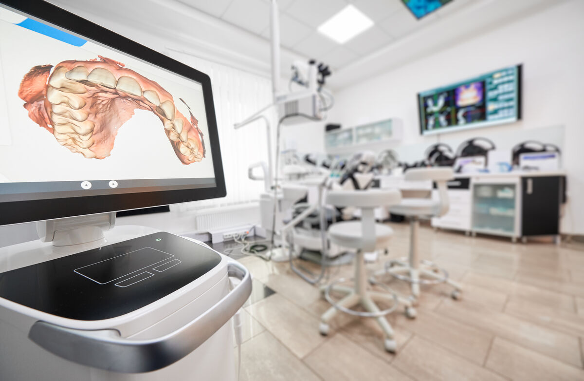
Cone Beam Computed Tomography is a specialized type of X-ray technology that captures a comprehensive, three-dimensional view of the dental and maxillofacial structures. Unlike a conventional panoramic or periapical X-ray, which flattens your anatomy into a single two-dimensional image, CBCT uses a cone-shaped beam of radiation that rotates around your head in a single sweep, typically taking between 200 and 600 individual projection images. Sophisticated software then reconstructs these projections into a volumetric dataset, essentially a digital cube of data that your dentist can explore in any plane: axial (top-down), sagittal (side-to-side), and coronal (front-to-back).
The result is an extraordinarily detailed model that reveals bone density, nerve canal locations, sinus anatomy, tooth root morphology, and even early pathological changes that would be invisible on a standard X-ray. In practical terms, this means your dentist can visualize the exact position of an impacted tooth relative to the inferior alveolar nerve, measure bone width and height at a planned implant site down to fractions of a millimeter, or identify a hairline root fracture that could otherwise go undetected for months.
"CBCT has done for dentistry what MRI did for orthopedics. We went from guessing at what lies beneath the surface to seeing it with remarkable clarity. Every implant I place today is planned in three dimensions before I ever pick up a surgical handpiece."
How the Technology Differs from Medical CT
It is important to distinguish dental CBCT from the large medical CT scanners you might encounter in a hospital. Medical CT scanners use a fan-shaped beam and require the patient to lie flat while the machine captures hundreds of thin cross-sectional slices. These machines are excellent for soft-tissue imaging but deliver a substantially higher radiation dose. Dental CBCT scanners, by contrast, are compact, allow the patient to sit or stand upright, complete the scan in under a minute, and deliver a radiation dose that is typically 5 to 20 times lower than a conventional medical CT. This dramatic reduction in dose, combined with superior resolution for hard-tissue structures, is what makes CBCT ideally suited for dental and maxillofacial applications.
Why Dentists Are Adopting CBCT at Record Rates in 2026
The adoption curve for CBCT has been remarkable. According to a 2025 survey by the American Dental Association, approximately 62% of dental practices in the United States now have an in-office CBCT unit, up from just 35% in 2020 and fewer than 10% in 2010. Several converging factors are driving this trend.
First, the cost of CBCT hardware has dropped significantly. Entry-level units that once cost $150,000 or more can now be purchased for under $70,000, with compact models designed for small practices available for around $50,000. Second, the reimbursement landscape has improved: more insurance carriers now recognize CBCT as a medically necessary diagnostic tool for specific indications such as implant planning, impacted teeth, and TMJ evaluation, rather than treating it as an optional upgrade. Third, the integration of AI-powered diagnostic software has made CBCT interpretation faster and more reliable, reducing the learning curve for general practitioners who previously relied on referrals to imaging centers.
Did You Know?
The latest generation of CBCT scanners can capture a full maxillofacial volume in as little as 5 seconds, reducing motion artifacts and making the technology suitable even for pediatric patients and those with anxiety disorders. Some 2026 models also include built-in cephalometric imaging, eliminating the need for a separate X-ray machine for orthodontic analysis.
Clinical Applications of CBCT Across Dental Specialties
The versatility of CBCT means it has become an indispensable tool across virtually every dental specialty. Here is how each field uses this technology in 2026.
Implant Dentistry
This remains the single largest application of dental CBCT. Before placing a dental implant, the surgeon must know the precise width, height, and density of the available bone, as well as the exact location of critical structures such as the inferior alveolar nerve, the mental foramen, and the maxillary sinus floor. CBCT data is imported into implant planning software, where the surgeon can virtually position the implant at the optimal angle and depth, then fabricate a surgical guide that transfers this plan to the patient's mouth with sub-millimeter accuracy. Studies published in 2025 show that guided surgery based on CBCT planning reduces implant placement errors by up to 74% compared to freehand placement.
Orthodontics
Orthodontists use CBCT to assess impacted canines, supernumerary teeth, root resorption risk, airway dimensions, and the precise three-dimensional relationship between teeth and bone. This data is increasingly combined with intraoral scan data to create comprehensive digital treatment plans for both traditional braces and clear aligner therapy.
Endodontics
Root canal specialists rely on CBCT to identify extra canals, detect periapical pathology that is not visible on two-dimensional radiographs, diagnose root fractures, and evaluate the outcome of previous root canal treatments. The American Association of Endodontists now recommends CBCT as a first-line diagnostic tool for complex endodontic cases.
Oral Surgery and Pathology
From impacted wisdom tooth extraction to the evaluation of jaw cysts, tumors, and fractures, CBCT provides the surgical team with a three-dimensional roadmap that reduces complication rates and improves surgical outcomes. CBCT is also used to evaluate TMJ disorders, assess condylar morphology, and plan orthognathic (jaw) surgery.
| Dental Specialty | Primary CBCT Applications | Benefit Over 2D X-Rays |
|---|---|---|
| Implant Dentistry | Bone volume measurement, nerve mapping, guided surgery planning | Up to 74% fewer placement errors |
| Orthodontics | Impacted tooth localization, airway analysis, root resorption assessment | True 3D tooth-root visualization |
| Endodontics | Extra canal detection, periapical lesion identification, fracture diagnosis | Detects up to 34% more lesions |
| Oral Surgery | Wisdom tooth nerve proximity, cyst evaluation, jaw fracture mapping | Reduced nerve injury rates |
| Periodontics | Bone defect characterization, furcation assessment, regenerative planning | Accurate 3D defect morphology |
CBCT vs. Traditional Dental X-Rays: Key Differences
Understanding the differences between CBCT and conventional dental radiographs helps patients appreciate when the more advanced imaging is truly necessary and when a simple X-ray is perfectly sufficient.
| Feature | Periapical / Bitewing X-Ray | Panoramic X-Ray | CBCT Scan |
|---|---|---|---|
| Dimension | 2D | 2D (flattened panorama) | Full 3D volume |
| Radiation Dose | ~5 microSv per image | ~15-25 microSv | ~30-200 microSv (field-dependent) |
| Scan Time | ~0.2 seconds | ~15 seconds | ~10-40 seconds |
| Bone Density Info | Limited | Minimal | Detailed, measurable |
| Nerve Canal Mapping | Not possible | Approximate | Precise, sub-millimeter |
| Typical Cost | $25-$50 per image | $80-$150 | $150-$600 |
It is worth emphasizing that CBCT is not a replacement for conventional X-rays. A standard set of bitewing radiographs remains the best tool for detecting interproximal cavities, while a periapical X-ray is often perfectly adequate for evaluating a single tooth's root and surrounding bone. CBCT is reserved for situations where three-dimensional information provides a clear diagnostic or treatment-planning advantage. This principle, known as ALARA (As Low As Reasonably Achievable), guides responsible use of all radiographic technology.
Important Consideration:
CBCT should only be prescribed when the clinical question cannot be adequately answered by a lower-dose conventional radiograph. Reputable practitioners follow the ALARA principle and will explain why a CBCT scan is specifically needed for your case. If a dentist routinely orders CBCT scans for every new patient exam without a specific clinical indication, you are within your rights to ask for the justification.
The Patient Experience: What to Expect During Your CBCT Scan
For patients who have never had a CBCT scan, the experience is straightforward and far less intimidating than a medical CT or MRI. Here is what typically happens.
You will be asked to remove any metal jewelry, eyeglasses, hearing aids, or removable dental appliances. The technician will position you in the scanner, which is an open, upright unit. Depending on the model, you will either sit in a chair or stand with your chin resting on a small support. A head stabilizer ensures you remain still during the scan. The machine's C-arm will rotate around your head in a single pass, taking approximately 10 to 40 seconds. During this time, you simply need to stay still and breathe normally. There is no noise beyond a quiet hum, no tunnel to enter, and no contrast dye to inject. The entire appointment, from check-in to completion, typically takes about 15 minutes.
The images are available almost immediately. Your dentist can view the 3D dataset on a computer screen within minutes of the scan, rotating and slicing through the volume in real time to identify areas of concern. Many practices now use large-screen monitors in the treatment room so that you can see your own anatomy and better understand your diagnosis and treatment options.
"Patients are often amazed when they see their own CBCT images on screen. Showing someone the exact position of their impacted wisdom tooth relative to their nerve, or the precise bone defect around a failing implant, transforms the conversation. It goes from abstract dental jargon to 'I can see it myself.' That understanding builds trust and helps patients make informed decisions."
Radiation Safety and Dose Comparison
Radiation exposure is understandably a concern for patients. It is helpful to put CBCT doses into context. The effective dose from a dental CBCT scan varies depending on the field of view (the area being imaged) and the specific machine settings, but it generally ranges from about 30 microSieverts for a small-field scan focused on a few teeth to about 200 microSieverts for a large-field scan of the entire maxillofacial complex.
To put this in perspective, the average American receives approximately 3,100 microSieverts of background radiation per year simply from natural sources such as cosmic rays, radon gas, and naturally occurring radioactive materials in soil and food. A single dental CBCT scan represents roughly 1 to 6% of your annual background exposure. A round-trip cross-country flight from New York to Los Angeles exposes you to about 40 microSieverts. A medical chest CT delivers approximately 7,000 microSieverts, which is 35 to 230 times more than a dental CBCT.
Radiation Dose in Perspective:
- Single dental periapical X-ray: ~5 microSv
- Full-mouth X-ray series (18 images): ~90 microSv
- Dental CBCT (small field): ~30-80 microSv
- Dental CBCT (large field): ~100-200 microSv
- Medical chest CT: ~7,000 microSv
- Annual US background radiation: ~3,100 microSv
That said, the principle of using the lowest possible dose for each patient remains paramount. Modern CBCT machines offer adjustable field-of-view settings, allowing the clinician to limit the scanned area to only the region of interest. Pediatric protocols with reduced exposure parameters are now standard on most 2026-model scanners. Pregnant women should avoid CBCT scans unless there is an urgent clinical need, and a lead thyroid collar is recommended for all patients.
Cost of a Dental CBCT Scan in 2026
The cost of a dental CBCT scan in 2026 varies based on geographic location, the size of the field of view, and whether the scan is performed at a dental office or an independent imaging center. Here is a general breakdown of what patients can expect to pay.
A small-field CBCT scan, which focuses on a specific area such as one or two teeth and the surrounding bone, typically costs between $150 and $300. A medium-field scan covering one full arch ranges from $250 to $400. A large-field or full-face CBCT, which captures both jaws, the sinuses, and the TMJs, generally costs between $350 and $600.
Insurance coverage for CBCT has improved considerably over the past few years. Many dental PPO plans now cover CBCT imaging when it is deemed medically necessary, particularly for implant planning, impacted teeth assessment, and TMJ evaluation. Coverage typically falls under the "diagnostic" benefit category, subject to your plan's annual maximum. Medical insurance may also cover CBCT scans ordered for pathology evaluation, sleep apnea assessment, or trauma-related imaging. Always obtain a pre-authorization and verify coverage with your carrier before the scan.
AI Integration: The Future of CBCT Diagnostics
Perhaps the most exciting development in CBCT technology in 2026 is the integration of artificial intelligence into the diagnostic workflow. Several FDA-cleared AI platforms now analyze CBCT datasets automatically, flagging potential pathologies such as periapical lesions, bone loss, cysts, airway constrictions, and even early-stage osteoporotic changes in the jaw. These tools do not replace the dentist's clinical judgment but serve as a powerful second opinion, catching findings that might be overlooked during a busy clinical day.
Leading AI platforms, including Overjet, Pearl, and Dentistry.AI, report detection sensitivities above 95% for common dental pathologies when analyzing CBCT data. The AI generates a structured report highlighting areas of concern, which the clinician then reviews and correlates with clinical findings. This workflow has been shown to reduce diagnostic errors by up to 40% and cut interpretation time in half, according to data published in the Journal of Dental Research in late 2025.
Beyond diagnosis, AI is also transforming treatment planning. Software now automatically segments the jawbone, identifies the nerve canal, and proposes optimal implant positions based on available bone volume and prosthetic requirements. What once took a specialist 45 minutes of manual planning can now be accomplished in under 10 minutes with AI assistance, with the clinician making final adjustments and approvals.
A Note on AI in Dentistry:
While AI-assisted diagnostics represent a significant advance, they are designed to augment, not replace, clinical expertise. The final diagnosis and treatment decisions always rest with the qualified dental professional. If your dentist uses AI-assisted CBCT analysis, this is generally a positive sign that the practice is committed to using the latest evidence-based tools for your care.
Sources
- American Dental Association. Survey of Dental Practice: Technology Adoption Trends 2025. ADA Health Policy Institute, 2025.
- Ludlow JB, Timothy R, Walker C, et al. Effective dose of dental CBCT -- a meta-analysis of published data and additional data for nine CBCT units. Dentomaxillofacial Radiology. 2024;53(2):138-149.
- Jacobs R, Salmon B, Codari M, Hassan B, Bornstein MM. Cone beam computed tomography in implant dentistry: recommendations for clinical use. BMC Oral Health. 2018;18(1):88.
- American Association of Endodontists. Joint Position Statement on the Use of CBCT in Endodontics: 2025 Update.
- Yilmaz E, Kayipmaz S, Tosun G. Artificial intelligence applications in dental CBCT analysis: a systematic review. Journal of Dental Research. 2025;104(11):1189-1200.
- U.S. Food and Drug Administration. Dental Cone-Beam Computed Tomography -- Information for Healthcare Providers. FDA.gov, updated 2025.
- National Council on Radiation Protection and Measurements. NCRP Report No. 184: Dental Diagnostic Imaging. NCRP, 2023.
FAQ: Your Top Questions About Dental CBCT Scans
Not at all. A CBCT scan is completely painless and non-invasive. You simply stand or sit still for 10 to 40 seconds while the machine rotates around your head. There is no need for injections, contrast dye, or any physical contact with the scanned area. The experience is similar to having a panoramic X-ray taken but provides vastly more detailed information.
As with any radiographic procedure, CBCT scans are generally avoided during pregnancy unless there is an urgent clinical need that outweighs the minimal radiation risk. If you are pregnant or suspect you may be, always inform your dentist before any imaging procedure. In most cases, non-urgent dental imaging can be safely postponed until after delivery.
Many dental insurance plans now cover CBCT imaging when it is medically necessary, particularly for implant planning, impacted teeth evaluation, and TMJ assessment. Coverage varies by plan, so it is always best to verify with your insurance carrier or ask your dental office to submit a pre-authorization. Some scans may also be covered under medical insurance if they are related to pathology, trauma, or airway evaluation.
CBCT scans are not routine and should only be taken when there is a specific clinical indication that requires three-dimensional imaging. Unlike regular dental checkup X-rays, there is no standard interval for repeat CBCT scans. Your dentist will recommend a new scan only when the diagnostic benefit clearly justifies the additional radiation exposure, such as evaluating healing after a bone graft or reassessing a previously treated lesion.
CBCT can reveal bone destruction, masses, and other changes in the jaw that may suggest a malignant or benign tumor. However, CBCT is not the primary screening tool for oral cancer. A clinical oral examination remains the first line of detection for soft-tissue cancers. If a suspicious lesion is found, CBCT may be used to assess bone involvement, and a biopsy will be required for a definitive diagnosis. Advanced soft-tissue tumors are typically evaluated with medical CT or MRI.