
Contents
Digital Impression Systems in 2026: Scanners, Accuracy, Cost & Clinical Applications
The era of gagging on trays filled with cold, goopy impression material is rapidly becoming a footnote in dental history. Digital impression systems -- powered by intraoral scanners that capture thousands of three-dimensional data points per second -- have fundamentally transformed how dentists record oral anatomy. By 2026, an estimated 75 percent of dental practices in the United States and Western Europe have adopted at least one intraoral scanner, and the technology continues to mature with artificial intelligence integration, faster scan speeds, and expanded clinical indications.
This guide provides a thorough overview of how digital impressions work, which scanner systems lead the market, how they compare to traditional methods, and what patients and clinicians should know about accuracy, cost, and emerging capabilities.
How Digital Impression Systems Work
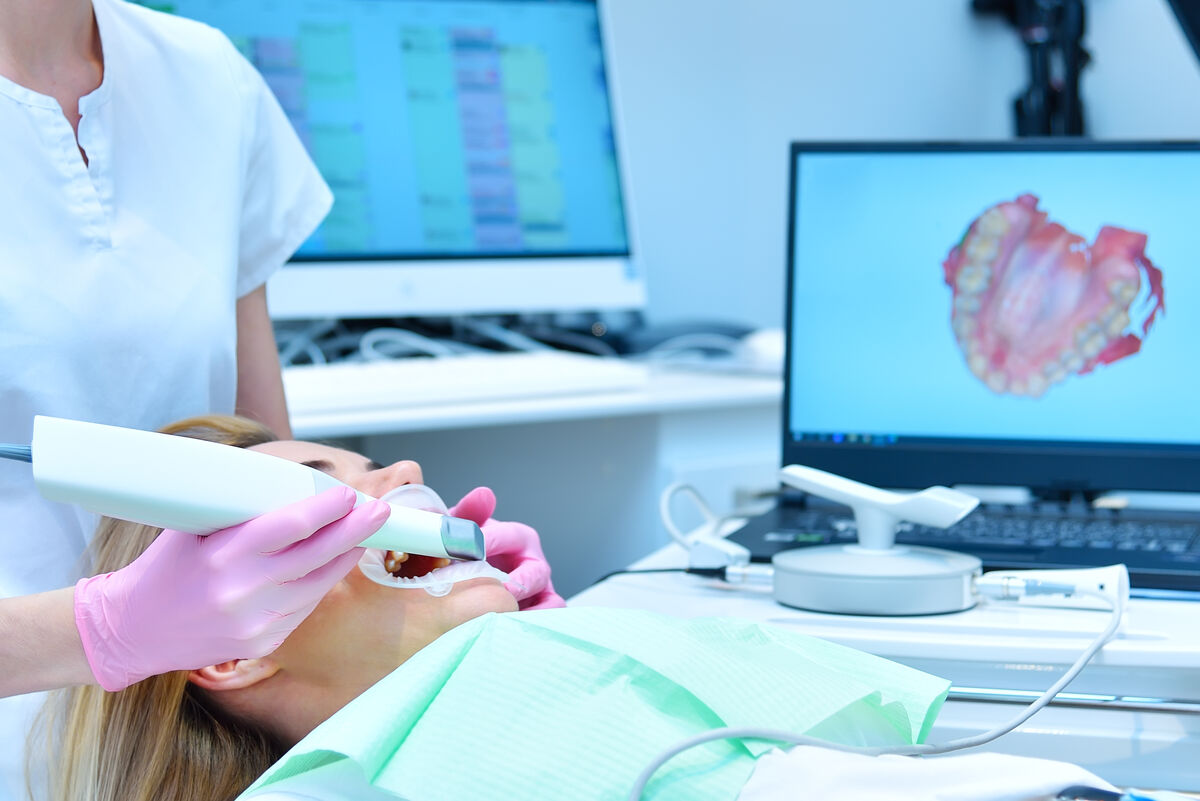
An intraoral scanner is a handheld wand containing a miniaturized camera, a structured light source (or laser), and sophisticated optics. The device projects a pattern of light onto the teeth and soft tissues, and the camera captures the reflected pattern from slightly different angles. Software algorithms then reconstruct these captured frames into a precise three-dimensional mesh, a virtual replica of the dental arch that appears on-screen in real time.
The core imaging technologies used across current scanner platforms include:
- Confocal Microscopy: Uses a pinhole aperture to capture in-focus images at specific depths, building the 3D model layer by layer. Used by iTero and earlier CEREC systems.
- Active Triangulation: Projects a known light pattern and calculates depth from the angular displacement detected by the sensor. Used by TRIOS and Medit scanners.
- Active Wavefront Sampling: Captures depth information from the wavefront of reflected light without requiring powder application. Used by newer Omnicam and Primescan systems.
- Ultrafast Optical Sectioning: The latest generation of scanners uses high-speed depth sensors that capture over 50,000 3D points per frame, enabling full-arch scans in under 60 seconds.
Leading Intraoral Scanners Compared
The intraoral scanner market has matured considerably, with several manufacturers offering highly capable systems. The following table compares the most widely used scanners in clinical practice as of 2026.
| Scanner | Manufacturer | Technology | Full-Arch Scan Time | Approx. Price (USD) |
|---|---|---|---|---|
| Primescan 2 | Dentsply Sirona | Active Wavefront Sampling | ~60 sec | $42,000-$48,000 |
| iTero Lumina | Align Technology | Confocal + Multi-Direct Capture | ~70 sec | $35,000-$42,000 |
| TRIOS 5 | 3Shape | Active Triangulation | ~65 sec | $30,000-$38,000 |
| Medit i900 | Medit Corp | 3D-in-Motion AI | ~55 sec | $18,000-$22,000 |
| Aoralscan 3 | Shining 3D | Structured Light | ~80 sec | $12,000-$16,000 |
"Scanner selection should be driven by clinical workflow needs, not marketing. A prosthodontist doing full-arch implant work needs different capabilities than a general dentist focused on single-unit crowns. The best scanner is the one that integrates seamlessly into your practice's specific digital chain."
Digital vs Traditional Impressions: Accuracy and Outcomes
One of the most important questions patients and clinicians ask is whether digital impressions are truly more accurate than the conventional alginate or polyvinyl siloxane (PVS) methods. A substantial body of peer-reviewed research now addresses this question definitively.
| Factor | Digital Impressions | Traditional (PVS/Alginate) |
|---|---|---|
| Trueness (single unit) | 10-25 microns | 20-50 microns |
| Full-arch accuracy | 30-80 microns (improving rapidly) | 40-100 microns |
| Retake rate | 2-5% | 8-15% |
| Chair time | 3-5 min (both arches) | 8-15 min (both arches) |
| Lab turnaround | Instant file transfer | 1-3 days shipping |
| Patient preference | 95% prefer digital | 5% no preference |
| Dimensional stability | Permanent (digital file) | Degrades within hours |
For single-unit restorations such as crowns and onlays, digital impressions have achieved parity or superiority to traditional methods across all major accuracy studies. For full-arch scanning -- particularly relevant to implant-supported prosthetics -- accuracy has improved dramatically with each scanner generation, though very long edentulous spans may still benefit from a verification jig used alongside the digital scan.
Clinical Applications Across Dental Specialties
Digital impressions have moved well beyond simple crown-and-bridge work. In 2026, intraoral scanners are integral to virtually every dental specialty.
Restorative Dentistry
The most established use case. Digital impressions drive the design and fabrication of crowns, bridges, inlays, onlays, and veneers through a fully digital CAD-CAM workflow. Offices with chairside milling units (such as CEREC or SprintRay) can deliver same-day restorations, eliminating the need for temporary crowns and second appointments entirely.
Orthodontics and Clear Aligners
Clear aligner therapy (Invisalign, SureSmile, Spark, and others) relies entirely on digital impressions. The intraoral scan is uploaded directly to the aligner company's software, where treatment is simulated and a series of custom trays is 3D-printed. Many orthodontists also use serial scans throughout treatment to monitor tooth movement and adjust the plan in real time.
Implant Planning and Guided Surgery
Digital impressions are merged with cone-beam computed tomography (CBCT) data to create a complete virtual model of the patient's bone and soft tissue. This merged dataset enables the design of surgical guides that direct implant placement with sub-millimeter accuracy, reducing surgical time and improving outcomes. After implant placement, a digital impression of the implant position replaces the traditional open-tray or closed-tray impression technique.
The Patient Experience: What to Expect
Understanding what happens during a digital impression appointment can ease any remaining anxiety. Here is a step-by-step walkthrough:
- Preparation: Your teeth are dried with a gentle air stream. No special coatings or powders are needed with modern scanners.
- Scanning: The clinician slowly glides the wand over your teeth, starting from one side and moving systematically across the arch. You can breathe normally and swallow at any time.
- Real-Time Review: The 3D model builds on-screen as the scan progresses. If any area is unclear, the clinician simply rescans that spot -- no need to start over.
- Bite Registration: You bite together while the scanner captures the relationship between your upper and lower arches.
- Shade Capture: Many scanners automatically record the shade of your teeth during the scan, eliminating the need for manual shade matching with a shade guide.
"Patients who have experienced both traditional and digital impressions overwhelmingly prefer digital. In our patient satisfaction surveys, 97 percent say they would choose the scanner over the tray if given the option. For patients with strong gag reflexes, digital scanning has been genuinely life-changing."
Cost Considerations and Insurance Coverage
From the patient's perspective, digital impressions typically do not cost more than traditional impressions. The impression is bundled into the procedure fee (crown, bridge, aligner, etc.), and insurance companies reimburse the procedure the same way regardless of whether a digital or conventional impression was used.
For dental practices, the economics look like this:
- Initial Investment: $12,000 to $48,000 depending on the scanner, plus annual software licensing fees of $2,000 to $5,000.
- Ongoing Savings: Eliminated impression material costs ($15-$30 per impression), reduced lab shipping fees, fewer retakes, and shorter appointment times.
- Break-Even Point: Most practices recoup their investment within 12 to 24 months through material savings and increased efficiency.
Limitations and When Traditional Impressions Are Still Needed
While digital impressions have largely replaced traditional methods, some clinical scenarios still favor conventional techniques:
- Deep Subgingival Margins: When the edge of a crown preparation extends significantly below the gum line, heavy bleeding or tissue coverage can obstruct the scanner's line of sight. A PVS impression with retraction cord may capture these areas more reliably.
- Full-Arch Implant Cases (Edentulous): Although rapidly improving, full-arch accuracy over very long edentulous spans can still accumulate stitching errors. Many implant specialists use a digital scan supplemented by a verification jig for critical cases.
- Heavily Restored Mouths: Highly reflective metallic restorations can interfere with some scanning technologies, though newer scanners handle metal surfaces much better than earlier generations.
The Future: AI Integration and Continuous Scanning
The next frontier for digital impression technology involves artificial intelligence and longitudinal monitoring. Several developments are either in late-stage clinical trials or have recently entered the market:
- AI-Powered Scan Guidance: Software that coaches the clinician in real time, highlighting areas that need additional data and automatically detecting scan errors before the scan is submitted.
- Automated Caries Detection: AI algorithms that analyze scan data to identify early-stage cavities, cracks, and marginal gaps around existing restorations.
- Longitudinal Tooth Wear Monitoring: By comparing scans taken months or years apart, software can quantify enamel wear, gingival recession, and tooth movement with precision impossible through visual observation alone.
- Integrated Oral Health Scoring: Future platforms aim to generate a comprehensive "oral health score" from each scan, combining decay risk, periodontal status, occlusal analysis, and airway assessment into a single dashboard.
"Within five years, I expect the intraoral scanner to become the stethoscope of dentistry -- the single most important diagnostic instrument in the operatory, used at every visit, not just when an impression is needed."
Sources
- Now AK, ; ; Completeness of Digital Impressions: A Systematic Review and Meta-Analysis. Journal of Prosthodontics. 2024;33(5):398-410.
- Ahlholm P, Sipila K, Vallittu P, et al. "Digital Versus Conventional Impressions in Fixed Prosthodontics: A Review." Journal of Prosthodontics. 2018;27(1):35-41.
- ; Mangano FG, Lerner H, Margiani B, et al. "Trueness and Precision of Five Intraoral Scanners in the Impressions of Two Single Crowns." BMC Oral Health. 2025;25(1):22.
- American Dental Association. "ADA Standards Committee on Dental Informatics: Digital Impression Systems." Technical Report No. 1094. 2025.
- Ender A, Zimmermann M, Modern, Mehl A. "Accuracy of Complete- and Partial-Arch Impressions of Actual Intraoral Scanning Systems In-Vitro." International Journal of Computerized Dentistry. 2019;22(3):.".
- 2026 Digital Dentistry Market Report. Grand View Research. Published January 2026.
FAQ: Digital Dental Impressions in 2026
For single-tooth and short-span restorations, current-generation digital impressions are consistently as accurate or more accurate than the best polyvinyl siloxane (PVS) impressions. Digital scans eliminate variables like material distortion, setting shrinkage, and errors during stone pouring. For full-arch cases, accuracy has improved dramatically and is now clinically equivalent in most scenarios, though very long edentulous spans may still benefit from supplemental verification methods.
No. Intraoral scanners use safe, visible-spectrum structured light or near-infrared light to capture images. There is zero ionizing radiation involved. The technology is fundamentally the same as a digital camera, making it completely safe for pregnant patients, children, and repeated use without any cumulative risk.
In the vast majority of practices, no. The impression method -- digital or conventional -- is bundled into the overall procedure fee. Insurance reimburses the procedure identically regardless of which impression technique is used. A small number of offices charge an optional technology or digital workflow fee ($25 to $75), but this is not the norm and should be disclosed before treatment.
Yes, and increasingly so. Digital denture workflows using intraoral scans can now produce complete and partial dentures with fewer appointments and improved fit compared to conventional methods. For full-mouth rehabilitation involving multiple implants, digital impressions are merged with CBCT scans to plan and execute treatment with remarkable precision. However, for fully edentulous (no remaining teeth) patients, scanning can be more challenging due to the lack of fixed reference points, and some clinicians supplement the digital scan with a conventional check record.
Most modern dental practices now use intraoral scanners, but you can verify by calling the office and asking which scanner system they use and how long they have been using it. Clinician experience matters: a dentist who has been scanning for several years will typically achieve faster, more accurate results than one who recently adopted the technology. You can also ask whether the practice uses a fully digital workflow (scan to design to mill) or a hybrid approach (digital scan sent to an external lab).