
Contents
Underbite Correction & Prognathism: Complete 2026 Treatment Guide
Prognathism -- commonly referred to as an underbite -- is a skeletal malocclusion in which the lower jaw extends beyond the upper jaw. While frequently viewed as a cosmetic concern, an untreated underbite can progressively damage teeth, compromise speech, strain the temporomandibular joint, and even interfere with breathing during sleep. In 2026, patients have access to a wider range of corrective strategies than ever before, from AI-guided orthodontic treatment planning to refined surgical protocols that dramatically reduce recovery times. This comprehensive guide walks you through every aspect of underbite diagnosis and correction so you can make a fully informed decision about your care.
What Is Mandibular Prognathism and How Is It Classified?
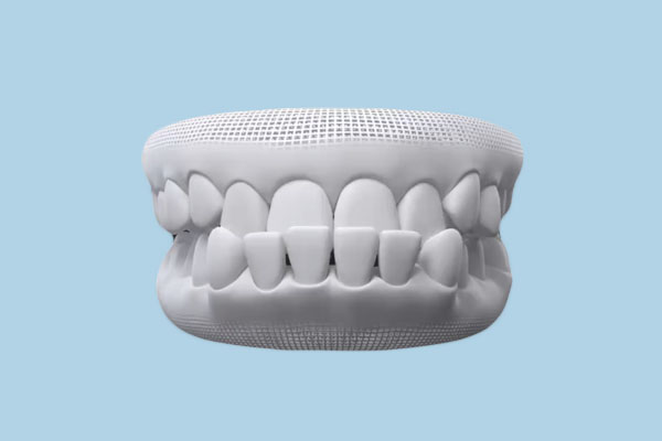
Mandibular prognathism is a dentofacial deformity classified as an Angle Class III malocclusion. In this condition the mandible (lower jaw) is positioned forward relative to the maxilla (upper jaw), causing the lower front teeth to sit ahead of the upper front teeth -- a relationship sometimes called an anterior crossbite. However, not every Class III case involves a large lower jaw. The discrepancy can stem from a normal-sized mandible paired with a deficient maxilla, or from a genuine overgrowth of the mandibular bone. Proper classification matters because it determines which jaw -- or both -- needs correction.
Understanding the Angle Classification System
Class I: Normal molar relationship with possible crowding or spacing. Class II: The lower jaw is positioned behind the upper jaw (overbite). Class III: The lower jaw extends in front of the upper jaw (underbite). Your orthodontist will use this system alongside cephalometric radiographs to pinpoint the exact skeletal discrepancy before proposing any treatment.
Severity is typically graded by measuring the overjet -- the horizontal distance between the upper and lower incisors. A negative overjet indicates the lower teeth are ahead of the upper teeth, and the magnitude of that negative value guides treatment selection.
| Severity Grade | Negative Overjet | Typical Treatment Pathway |
|---|---|---|
| Mild | -1 mm to -3 mm | Orthodontics alone (braces or clear aligners) |
| Moderate | -3 mm to -6 mm | Orthodontics with possible anchorage devices |
| Severe | Greater than -6 mm | Combined orthodontic-surgical approach |
Root Causes of Underbite Development
The etiology of prognathism is typically multifactorial, involving a combination of inherited skeletal patterns and environmental influences. Understanding these causes helps clinicians develop a targeted treatment strategy and, in some cases, allows parents to pursue early interceptive therapy for children who are genetically predisposed.
- Genetics and Hereditary Factors: The most dominant influence on jaw size and position is genetics. The Habsburg jaw, famously documented in the Spanish royal family, remains one of the best-known historical examples of inherited mandibular prognathism. If one or both parents have a Class III skeletal pattern, the likelihood of a child developing an underbite rises significantly.
- Childhood Oral Habits: Persistent thumb sucking, prolonged pacifier use beyond age three, tongue thrusting, and chronic mouth breathing can alter the trajectory of jaw development during the critical growth years. These habits exert sustained pressure on the developing bones and dental arches.
- Trauma and Injury: A fracture of the maxilla or mandible during childhood can disrupt normal growth plates, leading to asymmetric or disproportionate jaw growth as the child matures.
- Medical Conditions: Acromegaly (excess growth hormone), basal cell nevus syndrome (Gorlin syndrome), and certain craniofacial syndromes such as Crouzon syndrome or Apert syndrome can produce abnormal jaw dimensions.
- Premature Loss of Primary Teeth: Early loss of upper baby teeth -- without space maintenance -- can allow the lower arch to shift forward, sometimes mimicking or worsening a skeletal underbite.
"Early identification of Class III tendencies -- ideally by age seven -- gives orthodontists the greatest window of opportunity to redirect growth and potentially avoid surgical intervention later." -- American Association of Orthodontists, 2025 Clinical Guidelines
Health Consequences of an Untreated Underbite
Leaving a significant underbite untreated is not merely a cosmetic gamble. Over time, the skeletal imbalance creates a cascade of functional problems that worsen with age.
- Accelerated Enamel Erosion: Misaligned teeth make improper contact, grinding down enamel unevenly and exposing dentin. Studies show Class III patients lose enamel at 2 to 3 times the rate of patients with normal occlusion.
- TMJ Disorders (TMD): The abnormal bite forces the jaw joint into a strained position, often producing chronic pain, clicking, locking, and referred headaches.
- Speech Difficulties: Sibilant sounds (s, z, sh) and certain fricatives are commonly distorted because the tongue cannot make proper contact with the palate and teeth.
- Digestive Issues: Patients who cannot chew food thoroughly are at increased risk of gastrointestinal discomfort, acid reflux, and nutritional deficiencies.
- Obstructive Sleep Apnea (OSA): Severe maxillary deficiency narrows the upper airway, and emerging 2026 research links untreated Class III malocclusion with a 40% higher prevalence of mild to moderate OSA.
- Psychosocial Impact: Dissatisfaction with facial appearance frequently leads to reduced self-confidence, social withdrawal, and in adolescents, a measurably higher incidence of anxiety and depression.
Warning: Do Not Delay Evaluation
If your child consistently rests with the lower teeth in front of the upper teeth, or if you notice difficulty biting into food, schedule an orthodontic evaluation promptly. Class III problems rarely self-correct and tend to worsen during adolescent growth spurts.
Diagnostic Process and Severity Assessment
A thorough diagnosis of prognathism involves far more than a visual examination. In 2026, the standard diagnostic protocol combines clinical assessment with advanced imaging and digital analysis.
The orthodontist or oral surgeon will start with a comprehensive clinical examination, evaluating facial symmetry from the front and profile, assessing the dental midlines, measuring the overjet and overbite, and checking the molar relationship bilaterally. Next comes imaging: a lateral cephalometric radiograph is essential for performing a cephalometric analysis, which measures key skeletal angles such as the SNA (relationship of the maxilla to the skull base), SNB (relationship of the mandible to the skull base), and the ANB difference between them. A negative ANB angle confirms a skeletal Class III pattern.
Many practices now supplement this with a cone-beam computed tomography (CBCT) scan, which provides a three-dimensional view of the jaws, airway, and condyles. Digital intraoral scanning has also become routine, replacing traditional impressions and enabling clinicians to simulate treatment outcomes on screen before the patient commits to a plan.
"CBCT imaging has transformed our ability to visualize the precise three-dimensional relationship between the maxilla, mandible, and airway, enabling us to plan corrections with millimeter-level accuracy." -- Journal of Oral and Maxillofacial Surgery, 2025
Non-Surgical Treatment Options for Underbite Correction
Not every underbite requires surgery. When the skeletal discrepancy is mild to moderate -- or when the patient is young enough to benefit from growth modification -- non-surgical approaches can deliver excellent results.
Orthodontic Appliances and Early Intervention
Early interceptive treatment (Phase I orthodontics) targets children between the ages of 7 and 10, when the sutures of the maxilla are still responsive to orthopedic forces. The primary tools include:
- Rapid Palatal Expander (RPE): Widens a narrow upper jaw by applying lateral force to the midpalatal suture. This creates additional space in the upper arch and can improve the Class III relationship.
- Reverse-Pull Facemask (Protraction Headgear): Worn at night, this device hooks onto the upper molars and applies a forward-pulling force to the maxilla, stimulating forward growth. Compliance is critical, as the facemask must be worn 12 to 14 hours per day for optimal results.
- Miniscrew-Assisted Rapid Palatal Expansion (MARPE): A newer protocol that anchors the expander to temporary implant screws in the palate, allowing skeletal expansion even in older adolescents and some young adults whose sutures are beginning to fuse.
- Chin Cup Therapy: Less commonly used today, chin cups restrain forward mandibular growth in young children. Evidence for long-term effectiveness is mixed.
Clear Aligners for Mild Underbite Cases
For adults with a dental (non-skeletal) Class III pattern or a very mild skeletal discrepancy, clear aligners such as Invisalign can sometimes camouflage the underbite by retroclining the lower incisors and proclining the upper incisors. This approach has limitations -- it cannot move bone -- but it avoids both braces and surgery. In 2026, AI-powered treatment planning software has improved the predictability of aligner-based Class III camouflage, allowing orthodontists to identify suitable candidates more accurately than ever before.
Who Is a Good Candidate for Aligner Camouflage?
Ideal candidates have a negative overjet of no more than 3 mm, adequate bone support around the lower incisors, no significant facial asymmetry, and realistic expectations about the limitations of dental compensation. A detailed CBCT and cephalometric analysis is essential before committing to this pathway.
Orthognathic Surgery for Severe Prognathism
When the skeletal discrepancy is moderate to severe, orthognathic (jaw) surgery is the definitive treatment. This is a collaborative process involving both an orthodontist and an oral and maxillofacial surgeon. Treatment typically unfolds in three phases: pre-surgical orthodontics (12 to 18 months to decompensate the teeth and align each arch independently), the surgical procedure itself, and post-surgical orthodontics (6 to 9 months to refine the final bite).
The most common surgical procedures for Class III correction include:
- Bilateral Sagittal Split Osteotomy (BSSO): The mandible is split on both sides and repositioned posteriorly (set back). This is the most frequently performed procedure for mandibular prognathism.
- Le Fort I Osteotomy: The maxilla is separated from the skull base and advanced forward. This is used when the upper jaw is deficient rather than the lower jaw being excessively large.
- Bimaxillary Surgery: Both jaws are repositioned in the same operation. This is indicated for patients with discrepancies in both jaws and often produces the most dramatic improvement in facial balance.
Surgical Risks to Discuss With Your Surgeon
All surgery carries risks. The most significant concerns with orthognathic surgery include temporary or, rarely, permanent numbness of the lower lip and chin (inferior alveolar nerve damage), infection, relapse of the jaw position over time, unfavorable splits during mandibular osteotomy, and the need for revision surgery in roughly 3% to 5% of cases. Make sure your surgeon explains these risks thoroughly.
A major 2026 advancement is the widespread adoption of virtual surgical planning (VSP). Using the patient's CBCT data, surgeons can digitally simulate the osteotomies, reposition the virtual jaw segments to the optimal position, and have patient-specific cutting guides and fixation plates 3D-printed before the day of surgery. This has significantly reduced operative time and improved the accuracy of jaw repositioning.
Cost Breakdown and Insurance Coverage in 2026
Cost is one of the most significant factors influencing treatment decisions. Below is an overview of the typical costs patients can expect in the United States as of early 2026.
| Treatment Component | Estimated Cost Range (2026 USD) | Insurance Coverage Notes |
|---|---|---|
| Phase I Orthodontics (child) | $3,000 - $5,500 | Often covered partially under dental insurance |
| Full Orthodontics (braces) | $5,000 - $8,500 | Dental insurance may cover up to $2,000 lifetime max |
| Clear Aligners | $4,500 - $9,000 | Coverage varies; some plans treat same as braces |
| Orthognathic Surgery (single jaw) | $25,000 - $45,000 | Medical insurance often covers when medically necessary |
| Bimaxillary Surgery | $40,000 - $70,000 | Higher coverage likelihood with documented functional impairment |
| CBCT Scan | $250 - $600 | Typically covered as diagnostic imaging |
A critical distinction exists between dental insurance and medical insurance when it comes to underbite correction. Dental insurance typically covers orthodontics but has low annual and lifetime maximums. Medical insurance, on the other hand, may cover orthognathic surgery when it is deemed medically necessary -- meaning the condition causes functional impairment such as difficulty chewing, TMJ pain, or sleep apnea. Obtaining a letter of medical necessity from both your orthodontist and surgeon dramatically increases the chances of approval.
Recovery Timeline and Long-Term Outcomes
Understanding the recovery process is essential for setting realistic expectations, whether you are pursuing non-surgical or surgical correction.
For orthodontic-only treatment, patients should expect 18 to 30 months of active treatment followed by lifetime retainer wear (typically nightly for the first year, then several nights per week indefinitely). Discomfort is generally mild and manageable with over-the-counter analgesics.
For patients undergoing orthognathic surgery, the recovery timeline follows a predictable but demanding course. During the first two weeks, swelling peaks around days three through five, and patients are restricted to a liquid diet. Jaw immobilization with elastics is standard. By weeks three and four, patients typically transition to a soft-food diet and can return to sedentary work or school. From weeks six to eight, most patients resume normal eating, although hard or chewy foods should still be avoided. Full bone healing takes approximately 12 weeks, and residual numbness in the lower lip may persist for three to six months. The final orthodontic refinement phase then runs for another six to nine months.
Long-term studies tracking orthognathic surgery patients for 10 years or more show that skeletal relapse is minimal when rigid internal fixation is used and post-surgical orthodontics is completed properly. Patient satisfaction rates consistently exceed 90% in published outcome studies, with the most dramatic improvements reported in chewing efficiency, speech clarity, and self-confidence.
Tips for a Smoother Surgical Recovery
Stock your kitchen with broths, smoothies, protein shakes, and yogurt before surgery. Sleep with your head elevated on two pillows for the first two weeks. Apply ice packs to the cheeks for 20 minutes on, 20 minutes off during waking hours for the first 72 hours. Attend every scheduled follow-up appointment -- early detection of complications makes all the difference.
Sources
- American Association of Orthodontists -- Clinical Practice Guidelines for Class III Malocclusion, 2025 Update
- Journal of Oral and Maxillofacial Surgery -- Advances in Virtual Surgical Planning for Orthognathic Procedures, Vol. 83, 2025
- The Angle Orthodontist -- Long-Term Stability After Orthognathic Correction of Class III Malocclusion: A 10-Year Follow-Up Study, 2024
- American Journal of Orthodontics and Dentofacial Orthopedics -- MARPE Outcomes in Late Adolescents and Young Adults, 2025
- National Institute of Dental and Craniofacial Research -- Malocclusion and Orthodontic Treatment Data, 2025
FAQ: Underbite Correction and Prognathism
The American Association of Orthodontists recommends every child have an orthodontic screening by age seven. At this age, an orthodontist can identify early signs of a Class III skeletal pattern and begin interceptive treatment if necessary. Early intervention -- typically between ages 7 and 10 -- can guide jaw growth and significantly reduce the need for surgery in adulthood.
Clear aligners can only camouflage very mild underbites by tipping the teeth into a more favorable position. They cannot move jawbone. For moderate to severe skeletal underbites -- those with a negative overjet greater than about 3 mm -- orthognathic surgery combined with braces or aligners remains the standard of care. Using aligners alone in a case that truly requires surgery can lead to unstable results, root resorption, and worsening of the condition over time.
Most patients can return to work or school within two to three weeks, although swelling takes four to six weeks to largely resolve. A liquid diet is necessary for the first two weeks, transitioning to soft foods for another four weeks. Full bone healing occurs by approximately 12 weeks post-surgery. Residual numbness of the lower lip and chin is common and usually resolves within three to six months, though in rare cases it can persist longer.
Many medical insurance plans cover orthognathic surgery when it is deemed medically necessary -- meaning the underbite causes documented functional problems such as an inability to chew properly, TMJ disorders, speech impairment, or obstructive sleep apnea. The key is obtaining a detailed letter of medical necessity from both your orthodontist and your oral surgeon, supported by clinical records, imaging, and functional assessments. Pre-authorization is strongly recommended before proceeding.
Relapse is possible but uncommon with modern techniques. When rigid titanium fixation is used during surgery and post-surgical orthodontics is completed fully, long-term studies show that 90% to 95% of patients maintain stable results at the 10-year mark. The greatest risk factor for relapse is residual mandibular growth in patients who undergo surgery before skeletal maturity. For this reason, surgery is generally delayed until age 18 to 20 in males and 16 to 18 in females. Faithful retainer wear after orthodontics also plays a critical role in maintaining dental alignment.