
Contents
Early Cavity Detection in 2026: Signs, Diagnostic Technology, Treatment & Reversal
Dental caries -- the disease process behind cavities -- remains the most prevalent chronic disease worldwide, affecting 2.4 billion people according to the World Health Organization. Yet the most transformative shift in caries management over the past decade has been the move from "drill and fill" toward early detection and minimally invasive intervention. In 2026, dentists have access to diagnostic technologies that can identify demineralization months or even years before a visible cavity forms, opening the door to non-invasive treatments that can actually reverse the disease process and preserve natural tooth structure.
This comprehensive guide explains how cavities develop at the molecular level, the warning signs you can watch for at home, the advanced diagnostic tools your dentist uses in 2026, and the evidence-based strategies for preventing, arresting, and reversing early-stage caries without ever needing a drill.
Understanding Cavities: The Demineralization Process
A cavity is not an event; it is the end result of a dynamic, ongoing battle between demineralization (mineral loss) and remineralization (mineral repair) on the tooth surface. Understanding this process is key to understanding why early detection matters so much.
Here is how the process works: oral bacteria -- primarily Streptococcus mutans and Lactobacillus species -- metabolize sugars and starches from your diet, producing organic acids (primarily lactic acid) as a byproduct. These acids lower the pH at the tooth surface below the critical threshold of approximately 5.5, at which point calcium and phosphate ions begin dissolving out of the enamel's hydroxyapatite crystal structure. This is demineralization.
Between acid attacks, saliva acts as a natural repair system, buffering the pH back to neutral and supplying calcium, phosphate, and fluoride ions that reintegrate into the enamel lattice. This is remineralization. A cavity forms only when the cumulative balance tips toward demineralization over weeks and months -- meaning there were more and longer acid attacks than the saliva could repair.
"The paradigm has shifted entirely. We no longer view a cavity as something that simply 'happens.' It is the predictable outcome of a disease process that can be intercepted, managed, and in many cases reversed if we catch it early enough. The goal of modern caries management is to preserve tooth structure, not remove it."
The Five Stages of Cavity Progression
Cavities progress through predictable stages. Identifying which stage a lesion is in determines the appropriate treatment -- and whether reversal is still possible.
| Stage | What Is Happening | Reversible? | Treatment Approach |
|---|---|---|---|
| 1. White Spot Lesion | Subsurface enamel demineralization; surface intact but appears chalky white | Yes | Fluoride, CPP-ACP, resin infiltration |
| 2. Enamel Cavitation | Surface enamel breaks down; small hole or roughness forms | Partially (can be arrested) | Silver diamine fluoride, sealant, minimal filling |
| 3. Dentin Involvement | Decay penetrates through enamel into softer dentin; sensitivity begins | No | Conservative filling (composite or glass ionomer) |
| 4. Pulp Proximity | Decay approaches the nerve (pulp); sharp pain with hot/cold | No | Deep filling, indirect pulp cap, or root canal |
| 5. Abscess Formation | Bacteria infect the pulp; abscess forms at root tip | No | Root canal or extraction |
Early Warning Signs You Can Spot at Home
While many early cavities are invisible to the naked eye and require professional diagnostic tools, there are signs you can watch for between dental visits:
- White or chalky spots on teeth: These opaque patches, especially near the gum line or between teeth, are the hallmark of demineralization. They may appear matte compared to the surrounding shiny enamel.
- Increased sensitivity: A tooth that has become newly sensitive to cold drinks, sweets, or air may be experiencing enamel breakdown.
- Visible brown or dark spots: These may indicate arrested or progressing decay. Not all dark spots are active cavities, but they warrant professional evaluation.
- Floss catches or shreds: If dental floss consistently snags or frays at a specific contact point, it may be catching on a rough edge created by early decay between teeth.
- Food packing: If food consistently gets stuck in a particular area where it did not before, the tooth anatomy may have changed due to early cavity formation.
Modern Diagnostic Technologies for Early Detection
Beyond the traditional visual examination and dental radiographs (X-rays), dentists in 2026 have access to several advanced technologies specifically designed to detect cavities at the earliest possible stage.
Laser Fluorescence Devices
Devices such as DIAGNOdent (KaVo) use a diode laser that emits light at a specific wavelength (655 nm). When this light hits demineralized or bacterially infected tooth structure, the tissue fluoresces at a different wavelength than healthy enamel. The device assigns a numerical score (0 to 99) that correlates with the degree of demineralization, allowing the dentist to quantify the severity of a lesion and track changes over time.
Near-Infrared Transillumination
Near-infrared (NIR) transillumination shines safe near-infrared light through the tooth. Healthy enamel transmits the light, while areas of demineralization or decay absorb or scatter it, appearing as dark shadows on the captured image. This technology (used in devices like CariVu by Dentsply Sirona) is particularly effective for detecting interproximal (between-teeth) cavities without requiring X-ray exposure.
AI-Assisted Radiograph Analysis
Artificial intelligence algorithms trained on millions of dental radiographs can now identify caries lesions on bitewing X-rays with accuracy comparable to or exceeding experienced dentists. AI platforms such as Overjet, Pearl, and VideaHealth analyze each radiograph in seconds, highlighting suspicious areas and assigning confidence scores. A 2025 multicenter study published in the Journal of Dental Research found that AI-assisted detection increased interproximal caries identification by 25 percent compared to clinician-only evaluation.
| Technology | Best For | Radiation | Sensitivity | Limitations |
|---|---|---|---|---|
| Visual + Explorer | Obvious cavitation | None | Low for early lesions | Cannot detect subsurface lesions |
| Bitewing X-ray | Interproximal decay | Very low | Moderate | Misses ~30% of early enamel lesions |
| Laser Fluorescence | Occlusal (biting surface) decay | None | High | Can give false positives from staining |
| NIR Transillumination | Interproximal + crack detection | None | High | Limited on heavily restored teeth |
| AI-Assisted X-ray | All surfaces | Same as standard X-ray | Very High | Requires quality radiograph input |
"AI does not replace the dentist. It serves as a second set of eyes that never gets tired, never gets distracted, and processes every pixel of the image with the same consistency. In our practice, AI-assisted radiograph analysis has caught lesions we would have monitored for another year -- lesions that turned out to already involve dentin when we investigated."
Can Early Cavities Be Reversed Without Drilling
Yes -- and this is one of the most important developments in modern dentistry. When decay is caught at Stage 1 (white spot lesion, with the enamel surface still intact), several non-invasive treatments can halt and reverse the process:
- Prescription-Strength Fluoride: High-concentration fluoride varnish (22,600 ppm) applied in-office, combined with prescription fluoride toothpaste (5,000 ppm) for home use, drives mineral redeposition into the weakened enamel lattice. The resulting fluorapatite crystal is actually more acid-resistant than the original hydroxyapatite.
- Casein Phosphopeptide-Amorphous Calcium Phosphate (CPP-ACP): Products like MI Paste (GC America) deliver bioavailable calcium and phosphate directly to the tooth surface, providing the raw materials for remineralization. CPP-ACP with fluoride (MI Paste Plus) has shown superior remineralization in clinical trials.
- Resin Infiltration (Icon by DMG): A minimally invasive technique that fills the porous demineralized enamel with a low-viscosity resin without any drilling. The resin seals the lesion, blocks acid penetration, and restores the tooth's natural appearance by eliminating the white spot.
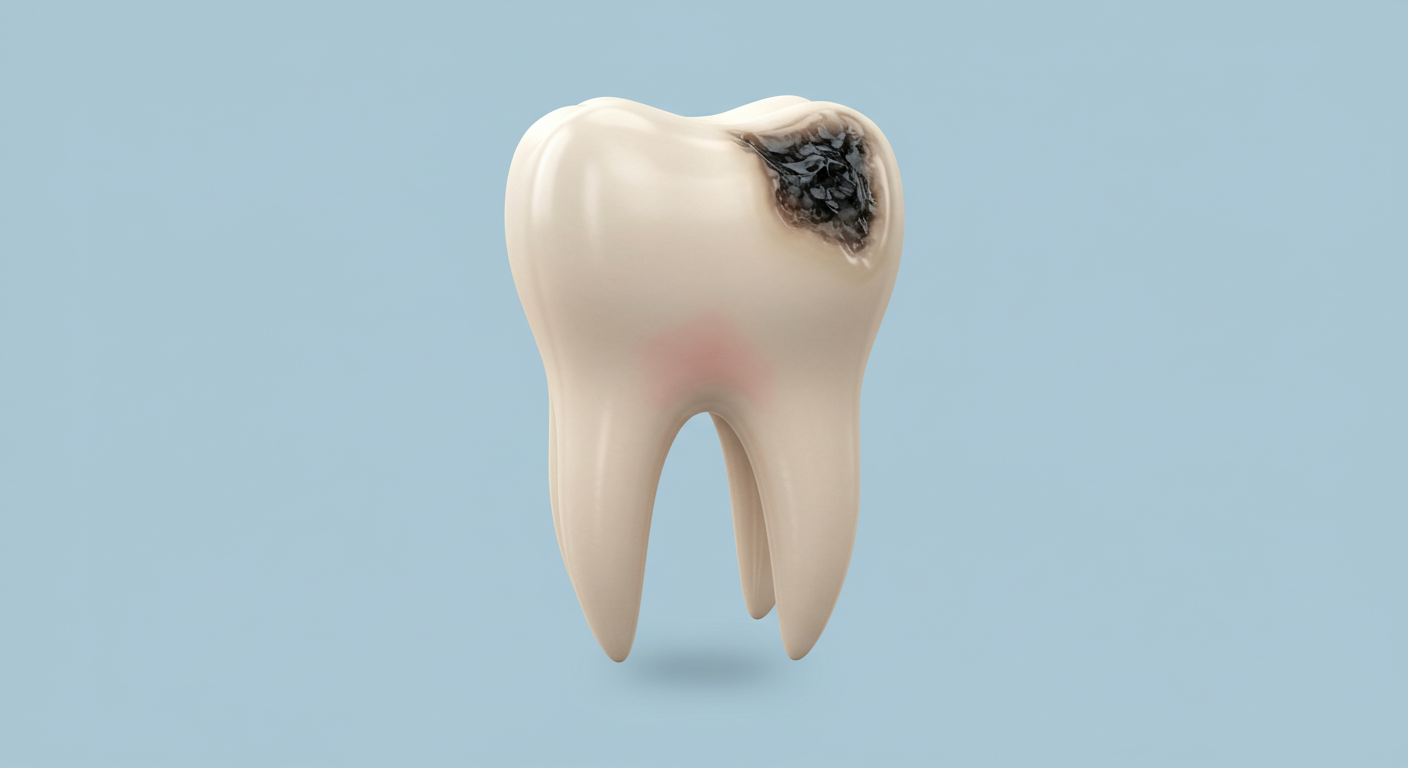
- Silver Diamine Fluoride (SDF): A liquid applied topically that kills caries-causing bacteria and hardens demineralized tooth structure. SDF is highly effective at arresting active cavities, though it leaves a permanent dark stain on treated areas, making it most suitable for primary teeth or non-visible surfaces.
Treatment Options by Cavity Stage
When a cavity progresses beyond the point of reversal, restorative treatment becomes necessary. The goal in 2026 is maximum conservation of healthy tooth structure.
- Small Enamel Cavities: Preventive resin restoration (sealant placed over the lesion) or micro-preparation with flowable composite resin. No anesthesia usually needed.
- Moderate Dentin Cavities: Conservative composite (tooth-colored) filling using minimally invasive preparation techniques. Air abrasion or laser preparation may replace the traditional drill in select cases.
- Large Dentin Cavities: Composite or ceramic inlay/onlay fabricated chairside using CAD-CAM technology for optimal fit and durability.
- Cavities Near the Nerve: Stepwise or selective caries removal, indirect pulp capping with bioceramics (Biodentine or MTA), and a final restoration. This approach preserves pulp vitality in many cases that previously would have required root canal treatment.
Prevention: Evidence-Based Strategies That Work
Prevention remains the most effective and least expensive approach to managing dental caries. The following strategies have the strongest evidence base:
- Fluoride Toothpaste (1,000-1,500 ppm): Brush twice daily for two minutes. Spit but do not rinse, allowing residual fluoride to continue working on the tooth surfaces.
- Limit Sugar Frequency: It is the frequency of sugar exposure, not the total amount, that drives caries. Five small sugary snacks throughout the day cause more damage than one large dessert, because each exposure triggers a 20 to 30 minute acid attack.
- Dental Sealants: Thin resin coatings applied to the grooves of permanent molars reduce cavity risk by 80 percent in sealed teeth. Recommended for children and adults with deep fissures.
- Regular Professional Cleanings and Exams: Every 6 months for average-risk patients, every 3 to 4 months for high-risk patients (dry mouth, history of frequent cavities, orthodontic appliances).
- Xylitol: Chewing xylitol gum (at least 6 grams daily) after meals has been shown to reduce S. mutans counts and lower caries incidence by 30 to 60 percent in clinical trials.
- Adequate Hydration and Saliva Flow: Saliva is the body's primary defense against cavities. Stay hydrated, and if you take medications that cause dry mouth, discuss saliva substitutes or stimulants with your dentist.
"If I could give one piece of advice to prevent cavities, it would be this: do not rinse after brushing. The single most impactful behavior change most patients can make is to spit out the toothpaste foam but leave the residual fluoride on their teeth. This simple habit change can reduce cavity formation by 25 percent."
Sources
- Fontana M, Zero DT. "Assessing Patients' Caries Risk." Journal of the American Dental Association. 2006;137(9):1231-1239.
- Pitts NB, Zero DT, Marsh PD, et al. "Dental Caries." Nature Reviews Disease Primers. 2017;3:17030.
- Schwendicke F, Frencken JE, Bjorndal L, et al. "Managing Carious Lesions: Consensus Recommendations on Carious Tissue Removal." Advances in Dental Research. 2016;28(2):58-67.
- Gomez J, Tellez M, Pretty IA, et al. "Non-Cavitated Carious Lesions Detection Methods: A Systematic Review." Community Dentistry and Oral Epidemiology. 2013;41(1):54-66.
- Cantu AG, Gehrung S, Dekker A, et al. "Detecting Caries Lesions of Different Radiographic Extension on Bitewings Using Deep Learning." Journal of Dental Research. 2025;104(1):78-86.
- American Dental Association Council on Scientific Affairs. "Caries Risk Assessment and Management." ADA Clinical Practice Guidelines. 2025.
- World Health Organization. "Global Oral Health Status Report." WHO Publications. 2022.
FAQ: Early Cavities and Detection
At the earliest stage -- the white spot lesion, where the enamel surface is still intact -- yes, the lesion can be remineralized and effectively "healed" with fluoride therapy, CPP-ACP products, and improved oral hygiene. However, once the surface enamel has physically broken down and a cavity (hole) has formed, the body cannot regenerate lost tooth structure. At that point, professional treatment is needed to stop the decay and restore the tooth. This is why catching cavities at the white spot stage is so critical.
The American Dental Association recommends that the frequency of dental X-rays be based on individual risk level, not a fixed schedule. For adults with low caries risk (no cavities in the past 3 years, good oral hygiene), bitewing X-rays every 24 to 36 months may be sufficient. For adults with elevated risk (history of frequent cavities, dry mouth, many existing restorations), bitewing X-rays every 6 to 12 months are appropriate. Your dentist will assess your personal risk factors and recommend a schedule tailored to your needs.
Not always. White spots can also be caused by fluorosis (excessive fluoride exposure during tooth development), enamel hypoplasia (developmental defects), or orthodontic decalcification (demineralization around brackets that has since been arrested). A dentist can differentiate between these causes using clinical examination and diagnostic tools like laser fluorescence. Active caries white spots typically have a chalky, matte texture and are located near the gum line, while fluorosis spots are usually symmetrical, well-defined, and present on both sides of the mouth.
SDF is safe and FDA-cleared for use in adults and children. It is highly effective at arresting active caries without drilling. However, SDF does permanently stain the treated decayed area dark brown or black through a chemical reaction between the silver and the decayed tooth protein. It does not stain healthy enamel. For this reason, SDF is most commonly used on primary (baby) teeth, root surface cavities in elderly patients, or non-visible surfaces where aesthetics are not a primary concern. When used on visible surfaces, the stained area can later be covered with a tooth-colored filling material (the SMART technique).